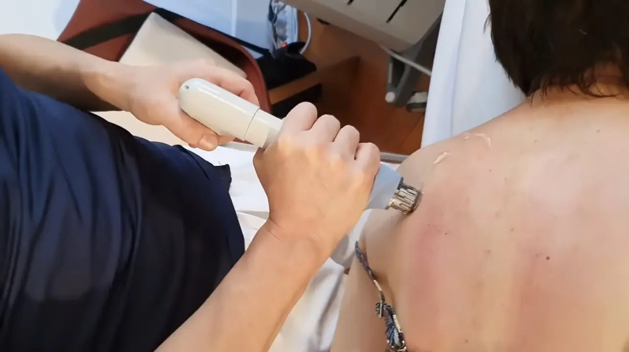
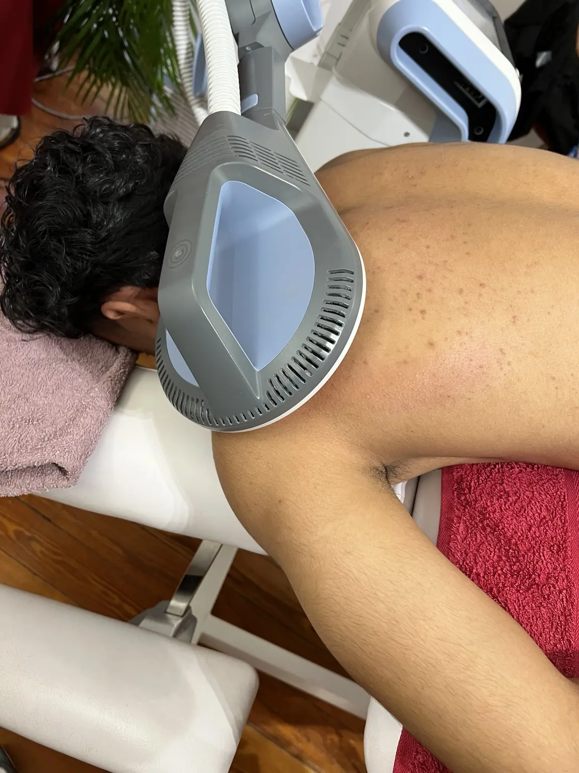
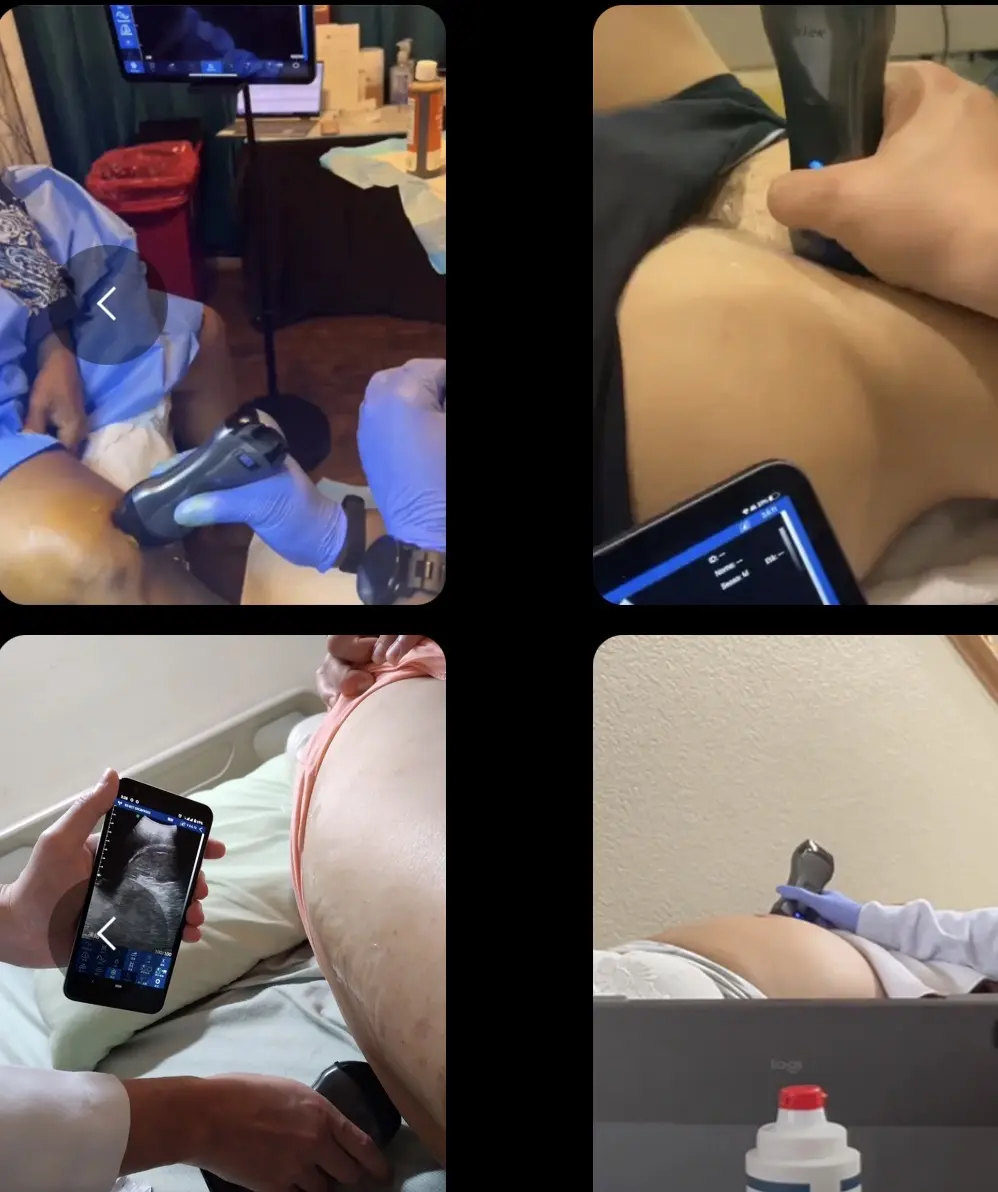
Structure
Accessible appearance of selected muscles, tendons, bursae and soft tissues.
 FR
FRDynamic ultrasound · Geneva
Real-time imaging of muscles, tendons, superficial joints and soft tissues, used as an adjunct to history and functional examination.
Rue de Fribourg 7 · 1201 Geneva
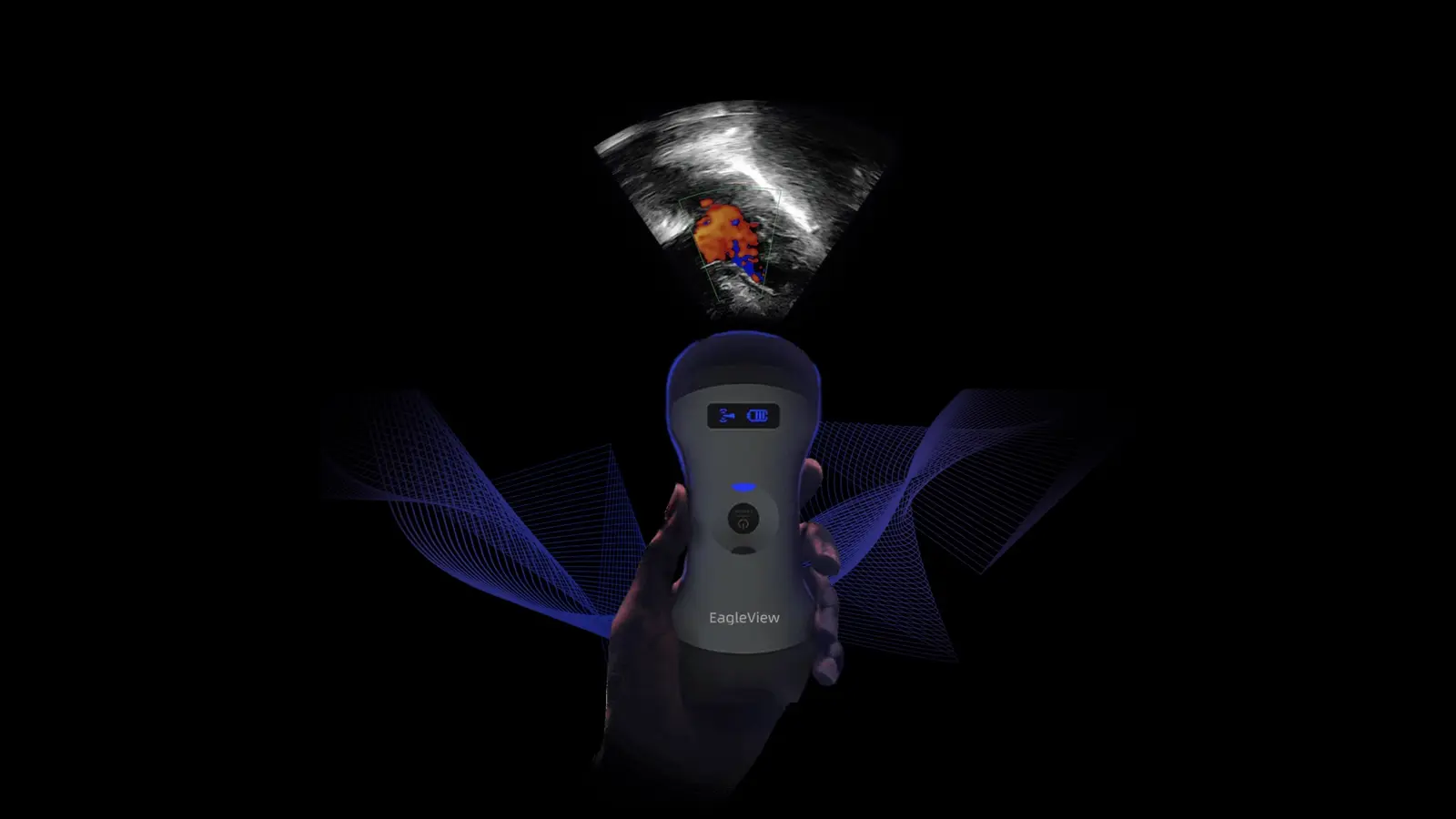
Musculoskeletal ultrasound can examine selected structures in real time, compare the symptomatic side with the opposite side and observe how a tendon or muscle behaves during a chosen movement.
Imaging is never interpreted alone. Findings may exist without explaining symptoms, while significant pain can occur without visible tissue damage. History, function and clinical tests remain essential.
When the question exceeds the capabilities of in-clinic ultrasound, radiology imaging or specialist medical review may be recommended.
02 · IMG CLINICAL REASONING
The clinical question determines the area, position and movement assessed.
Accessible appearance of selected muscles, tendons, bursae and soft tissues.
Gliding, contraction, continuity and relationships during movement.
Differences between the symptomatic and opposite side when comparison is useful.
Selected images that may support the clinical record and follow-up.
03 · CLINICAL PATHWAY
The examination is non-invasive and does not use ionising radiation.
Define the structure or movement to examine.
Place the region comfortably and apply gel to the skin.
Move the probe and reproduce relevant movements.
Relate images to symptoms, tests and the method’s limitations.
04 · INDICATIONS
05 · FAQ
It is normally painless. Light pressure may feel tender over an already sensitive area.
No. Ultrasound uses sound waves, not ionising radiation.
Selected images may be included in the clinical record depending on the question and follow-up.
No. It may clarify selected findings but must be interpreted with history, examination and sometimes other investigations.
06 · EXPLORE
Continue with the topics most directly connected to this assessment.
Your next step
Contact IMG Clinic directly or complete the Clinical Pathway Engine™ beta to prepare an initial clinician-reviewed pathway.